Anatomy of the Foot and Ankle Astoria Foot and Ankle Surgery
Dr. Ebraheim's educational animated video describes anatomical structures of the foot and ankle, The Bony Anatomy, The Joints, Ligaments, and the Compartment.

Ankle and Foot Pain Massage Therapy Connections
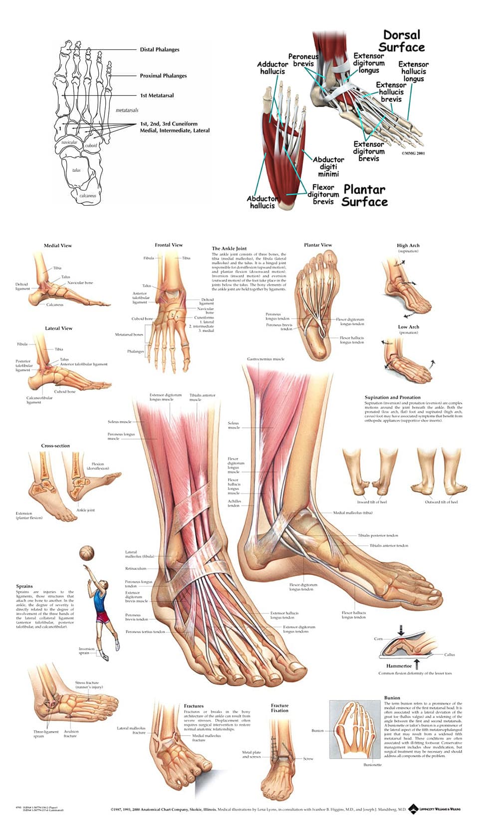
Bones of foot. The 26 bones of the foot consist of eight distinct types, including the tarsals, metatarsals, phalanges, cuneiforms, talus, navicular, and cuboid bones. The skeletal structure of.

Loading... Human anatomy chart, Foot anatomy, Nerve anatomy
The foot (pl.: feet) is an anatomical structure found in many vertebrates.It is the terminal portion of a limb which bears weight and allows locomotion.In many animals with feet, the foot is a separate [clarification needed] organ at the terminal part of the leg made up of one or more segments or bones, generally including claws and/or nails.

Foot Anatomy and Function पाद pāda Elliot's WebSite
Tarsals. The tarsals are a group of seven bones close to the ankle. The proximal tarsal bones are the talus and the calcaneus, which is the largest bone of the foot. The talus is on top of the.

Foot Anatomy Muscles Foot Archives Anatomy Human Body Foot anatomy
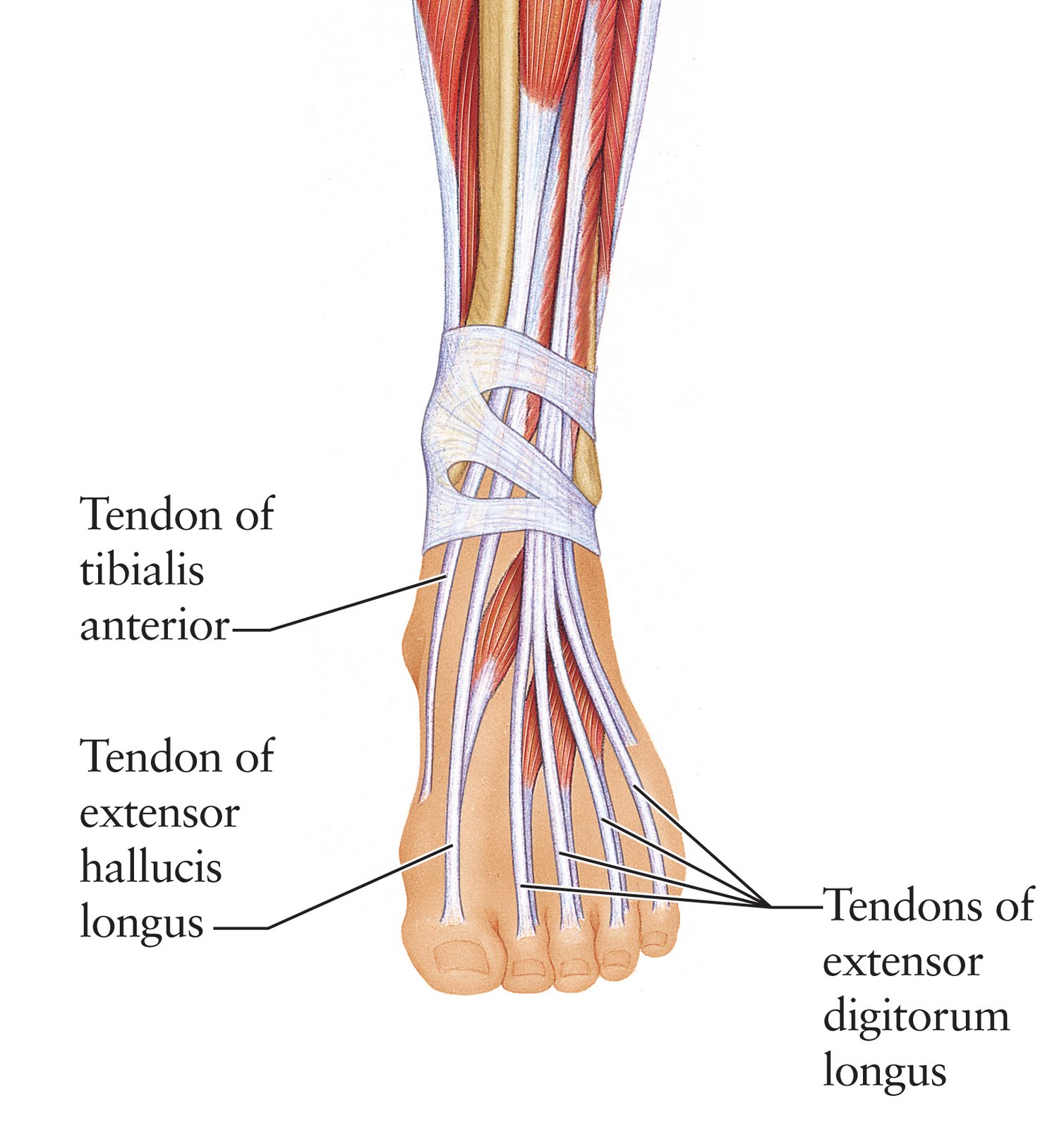
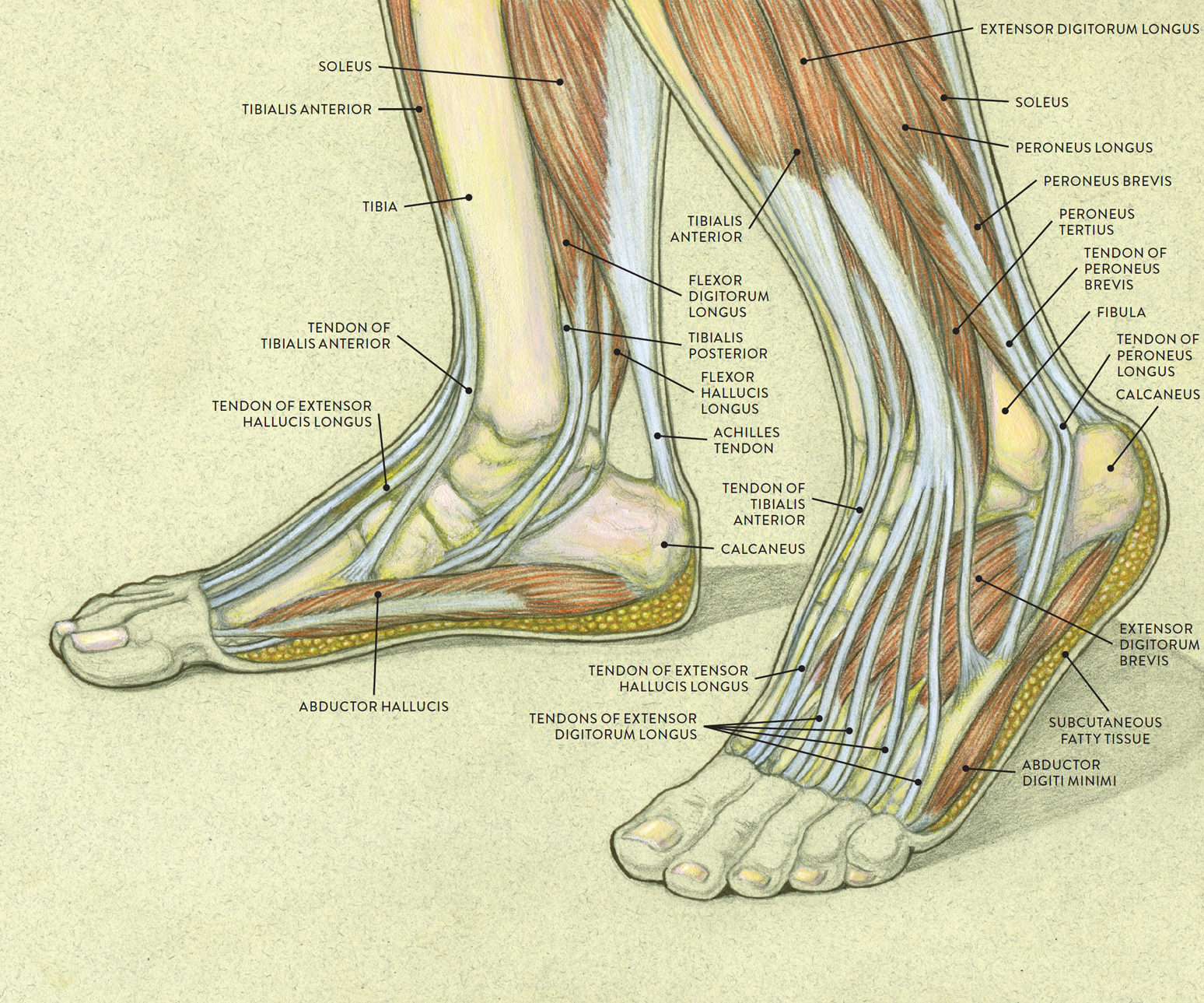
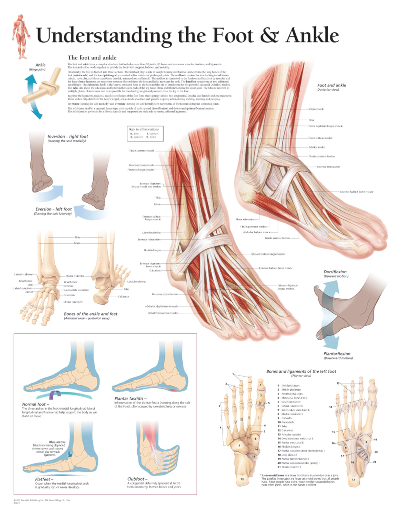
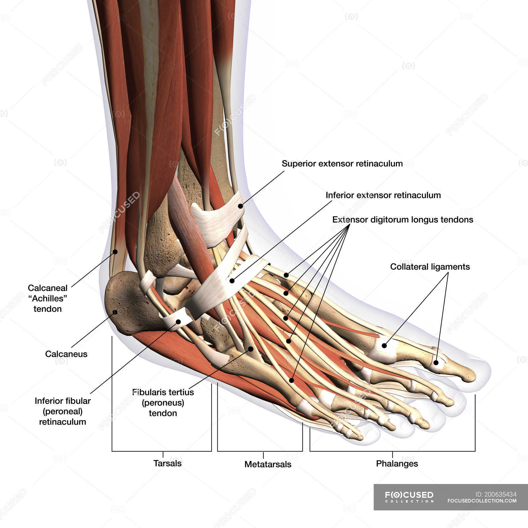
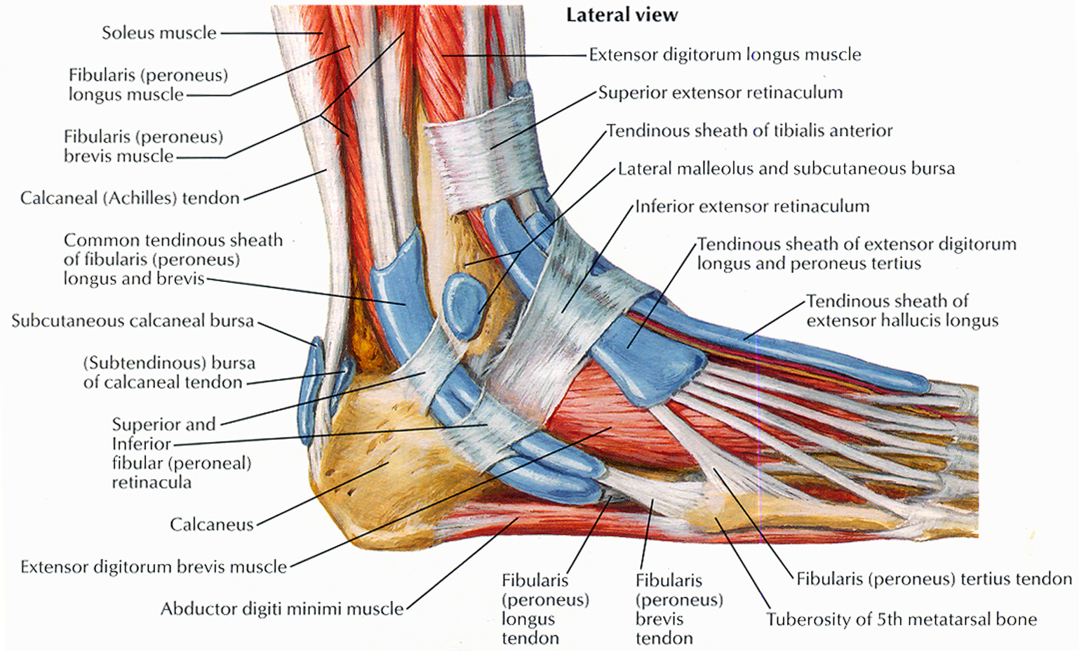
Foot and ankle anatomy consists of 33 bones, 26 joints and over a hundred muscles, ligaments and tendons. This complex network of structures fit and work together to bear weight, allow movement and provide a stable base for us to stand and move on. The foot needs to be strong and stable to support us, yet flexible to allow all sorts of complex.

Diagram Of A Human Foot Human Foot Diagram Anatomy Organ Anatomy
Ankle anatomy. The ankle joint, also known as the talocrural joint, allows dorsiflexion and plantar flexion of the foot. It is made up of three joints: upper ankle joint (tibiotarsal), talocalcaneonavicular, and subtalar joints.The last two together are called the lower ankle joint. The upper ankle joint is formed by the inferior surfaces of tibia and fibula, and the superior surface of talus.
Human Anatomy for the Artist The Dorsal Foot How Do I Love Thee? Let
Gastrocnemius (calf muscle): One of the large muscles of the leg, it connects to the heel. It flexes and extends the foot, ankle, and knee. Plantaris: This small, thin muscle is absent in about.

anatomy of the foot Ballet News Straight from the stage bringing
Foot. The foot is the lowermost point of the human leg. The foot's shape, along with the body's natural balance-keeping systems, make humans capable of not only walking, but also running.
Muscles of the Leg and Foot Classic Human Anatomy in Motion The
The anatomy of the foot. The foot contains a lot of moving parts - 26 bones, 33 joints and over 100 ligaments. The foot is divided into three sections - the forefoot, the midfoot and the hindfoot. The forefoot. This consists of five long bones (metatarsal bones) and five shorter bones that form the base of the toes (phalanges).

Toe Dislocation JOI Jacksonville Orthopaedic Institute
Use these bones of the foot quizzes to master your identification skills. Overview of the bones of the foot and their divisions into the hindfoot, midfoot and forefoot. With a total of 26 bones in each foot, learning the bony anatomy of the foot is no piece of cake. That is, the memorization aspect.

image lateral_ankle for term side of card Ligament Tear, Ligaments And
The feet support the human body when standing, walking, running, and more. They are complex structures with 26 bones. Learn more about foot bones and foot anatomy here.

hand bone and tendon chart Artist The Dorsal Foot How Do I Love
The foot is a part of vertebrate anatomy which serves the purpose of supporting the animal's weight and allowing for locomotion on land. In humans, the foot is one of the most complex structures in the body. It is made up of over 100 moving parts - bones, muscles, tendons, and ligaments designed to allow the foot to balance the body's.
Understanding the Foot & Ankle Scientific Publishing
Cuboid: This multi-faceted bone sits on the outside of the foot near the fifth phalanx (little toe). Cuneiforms: These three small bones are closest to the five metatarsal bones. They sit in a row.
Anatomy of human foot with labels on white background — ankle, leg
The foot is a complex anatomic structure composed of numerous bones, joints, ligaments, muscles, and tendons responsible for the complex coordinated movements of gait and our ability to stand upright. By definition, the foot is the lower extremity distal to the ankle joint. The ankle joint (sometimes referred to as the tibiotalar joint) is the result of the assembly of the talus and the recess.
Foot Anatomy Bones, Muscles, Tendons & Ligaments
The Anatomy of Feet: Bones and Structure. The foot is composed of 26 bones, making up about one-quarter of all the bones in the human body. These bones are divided into three main regions: the hindfoot, midfoot, and forefoot. The hind foot consists of the talus and calcaneus bones, which form the ankle joint and provide stability for weight.

Tendon Diagram Leg muscles leg tendons hamstrings diagram
Ligaments of the Foot and Ankle. Tell us where the pain is. Use our interactive tool. Use our Anatomy tools to learn about bones, joints, ligaments, and muscles of the foot and ankle. FootEducation is committed to helping educate patients about foot and ankle conditions by providing high quality, accurate, and easy to understand information.